Cardiac Catheterization
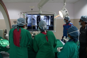
Cardiac catheterization is a common diagnostic test performed to evaluate the condition of heart muscle, valves, and vessels.
- During this procedure, special long, flexible tubes, called angiography catheters, will be inserted into your heart and coronary arteries.
- Contrast media (also called dye) is injected through the angiography catheter while x-ray images are taken.
- The dye causes areas where blood flows, including vessels and heart chambers, to temporarily become darker than the surrounding tissue. This enables the physician to see how effectively your heart is pumping, and to determine if there are any narrowed blood vessels
- Blood pressure measurements are also taken at this time.
Unlike cardiac surgery, most of the cardiology procedures have common steps with the changes being in the selection of “the interventional triad”: the Guiding Catheter, the Guide Wire and the Balloon (depending on the procedure). The basic process of cardiac catheterization remains the same.
Diagnostics and procedures in Cardiology may be divided into two broad categories, arrhythmia related procedures and anatomy related procedures.
Anatomy related diagnosis and treatment
Percutaneous Transluminal Mitral Commissurotomy PTMC
PTMC is a procedure where a balloon is used to dilate a stenosed mitral valve by threading it onto a catheter that is maneuvered into the mitral valve site. The most beneficial and effective technique was developed by Inoue in 1982.
PDA coil closure
A steel or MRI compatible metal coil is placed across the PDA to help In thrombotic closure – Thrombus (Clot) formation occurs on the metal coil and the PDA is sealed.
Pulmonary Valve Balloon Dilatation PVBD
This is done for Pulmonary valvular stenosis. A balloon catheter is maneuvered into the pulmonary valve through a right heart catheterization procedure and is inflated (valve is dilated) in a single stage.
Aortic Valve Balloon Dilatation AVBD
This is done for Aortic valvular Stenosis. A balloon catheter is maneuvered into the pulmonary valve through a left heart catheterization procedure and is inflated (valve is dilated) in a single stage.
Coarctation of Aorta Dilatation
When there is a block in the Aorta after the arch (thoracic or abdominal segments) obstructing the flow to the lower limbs of the body, a balloon is placed across this obstruction and inflated clearing the way for the blood to flow.
Peripheral Angiogram
The standard angiogram procedure is performed for other vascular structures of the body under investigation for obstructions/anomalies.
Pericardial Effusion Tapping (PE Tapping)
A catheter is inserted through the intercostal spaces (between the ribs) into the pericardial sac and pre-calculated (excess) pericardial fluid is aspirated (removed) using a syringe. The position of catheter confirmed using fluoroscopy and the liquid level is assessed using echocardiography.
Percutaneous Transluminal Angioplasty (PTA)
This is a generic name given to the interventional procedure that follows the angiogram. Angioplasty attempts to bring the vasculature back to normal through different means (stent, removal of plaque…). This procedure can be done anywhere in the body and the name changes according to the location such as: Coronary, Renal, Abdominal, Sub-Clavian, Carotid, Iliac, Brachial, Femoral, etc.
SAM Dilatation
More of a palliative procedure. A Sub Aortic Membrane, which is absent in a normal heart, causes obstruction to blood flow out of the Left Ventricle into the aorta. A balloon is inflated in the membrane area to suppress the growth so as to improve blood flow. It is generally considered not very effective. Surgery is the final option.
Tricuspid Valve Balloon Dilatation (TVBD)
Procedure similar to PTMC but the tricuspid valve is involved.
Balloon Atrial Septostomy (BAS)
This is a unique procedure to enable the mixing of pure and impure blood which enhances survival of the patient. This is normally done within one week to three months of birth generally for DTGA situations.
Arrhythmia related diagnosis and treatment
Electro-Physiological Study (E-P Study):
The normal heart beats according to the electrical impulses from the sinoartial node to the atrioventricular node. Sometimes additional abnormal path ways form and cause dangerous changes to the normal rhythm of the heart (arrhythmias) EP study is a diagnostic procedure done to find out extra pathways that cause these arrhythmias.
Radio Frequency Ablation (RF Ablation):
This is a remedial procedure for arrhythmias. A special catheter is maneuvered into the position identified by the EP study to be the anomalous pathway and the tip of the catheter is heated through application of Radio Frequency and burn the extra pathways preventing further arrhythmias (ablated).
Temporary Pacemaker Implantation (TPI):
A normal heart beats at a “pace” that causes the heart to eject blood into the aorta at the required pressure to circulate to the whole body. Due to various reasons the heart begins to slowdown leading to a condition called Bradycardia. Consequently the blood flow to the systemic circulation reduces (low cardiac output) leading to other complications. Temporary Pacemaker Implantation is a procedure wherein a catheter is positioned into the heart muscle and electrical impulses sent from an external pacemaker at the required pace to enable the heart to pump out its normal volume. This can be done under situations ranging from emergency interventions (accidents, organ failures, etc.) to procedural requirements in the Cath lab. In an emergency situation the TPI may itself suffice till the necessary action is taken and the heart returns to normal. Cath procedures requiring TPI include the process of Permanent Pacemaker Implantation (PPI), TPI is done to keep the heart beating normally till the PPI is completed.
Permanent Pacemaker Implantation (PPI):
When a TPI fails or the heart muscle is beyond salvage and cannot regain its original pace a PPI is performed. This is a semi surgical procedure done in the Cath lab itself. Pacemaker is a little device implanted in the chest to regulate the heart rate and rhythm. Through a simple procedure the implantation is often done in the operation theatre or cardiac catheterization lab, and usually takes an hour or two.
Pacemakers usually are implanted under local anesthesia, one will be relaxed, but awake, during surgery. Usually, pacemakers are implanted just under the skin in the upper chest. Typical complications for pacemaker implants are not life threatening, but may require a repeat operation or a longer than normal hospital stay. Common complications include bleeding, infection, lead dislodgment, and lead or pacemaker problems following surgery. Complications occur less than 1% of the time.
Intra Cardiac Defibrillator Implantation:
When the ventricles of the heart especially the left one beats on its own at a fast rate eg: 200bpm as against the normal 120bpm, it is called Ventricular Tachycardia. When this arrhythmia cannot be cured by ablation, the only other solution is to apply an electrical shock to bring it back to normal rhythm. Patients are implanted with a defibrillator that is programmed to sense this VT and “shock” it back to normal. The procedure for implantation is similar to that of the pacemaker but the instrument is a defibrillator instead of pacemaker.
Biventricular Pacemaker Implantation:
The left and right ventricles of a healthy heart pump out blood in a synchronized beat /rhythm. Some hearts do not have this synchronization, which leads to cardiac dysfunction and heart failure. To help such conditions, a special variety of pacemaker is implanted to bring the two ventricles back into sync and consequently improving the functioning of the heart.
Coronary related diagnosis and treatment
Coronary Angiogram
Angiography (also called cardiac catheterization or a heart cath) is a common diagnostic test performed to evaluate the condition of heart muscle, valves, and vessels. This procedure is most commonly performed to determine a patient’s cardiac condition and what form of treatment is required, including: medical management, angioplasty (PTCA, stenting or balloon widening of a vessel), or surgery.
Percutaneous Transluminal Coronary Angioplasty PTCA
This is a procedure in which a balloon is used to dilate blocked coronary arteries. This procedure has its advantages in that it postpones or avoids CABG surgery, but is limited to the extent of blockage. The process of Atherosclerosis may be far to advanced in certain cases for the procedure to successfully restore blood flow. In such cases CABG /OPCAB are the available surgical options.
Percutaneous Transluminal Angioplasty (PTA)
This is a generic name given to the interventional procedure that follows the angiogram. Angioplasty attempts to bring the vasculature back to normal through different means (stent, removal of plaque…). This procedure can be done anywhere in the body and the name changes according to the location such as: Coronary, Renal, Abdominal, Sub-Clavian, Carotid, Iliac, Brachial, Femoral, etc.
Percutaneous Trans-Septal Myocardial Ablation (PTSMA)
Each chamber of the heart has an optimal filling volume, which is in exact proportion to the other chamber volumes. When the Left Ventricular Volume is reduced, the carrying capacity/pumping volume also reduces, consequently the patient suffers secondary complications. This reduction in volume is sometimes due to Septal thickening. PTMA involves injecting an alcohol into the vessel supplying the thickened vessel dies part becomes thin. LV volume increases.


